
1. Background:
2. Benefits of cardiac MRI use:
Cardiac MRI is well suited for the examination of mitral regurgitation because it can accurately quantify the regurgitant volume and left ventricular volume. It is totally non-invasive and does not require contrast.
3. MRI Technique:
Short and long axis SSFP images are obtained to quantify end-diastolic and end-systolic LV volumes from which one can calculate the ejection fraction.
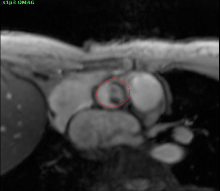
Phase-contrast images are obtained perpendicular to the aorta and/or the main pulmonary artery just distal to the respective valves.

click to enlarge
For accurate results, an identically prescribed phase-contrast image of a stationary phantom is used for baseline correction of the flow data.
4. Analysis:
The stroke volume is determined from the short axis SSFP images.
SV = LV EDV – LV ESV
The forward flow is measured using the phase-contrast images.
Phase Contrast Images

MR regurgitant volume = SV – FF
Regurgitant fraction = MR regurgitant volume / LV Stroke Volume
Using the short and long axis SSFP views, the anatomy of mitral valve and the direction of the regurgitant jet can also be assessed
5. Which imaging findings affect treatment?
The severity of mitral regurgitation can be quantified in terms of regurgitant volume or regurgitation fraction. Surgery is generally reserved for patients with severe mitral regurgitation unless they are going for cardiac surgery for other reasons. Surgical timing generally depends on symptoms, LV ejection fraction, and LV dimensions. AHA guidelines are shown in detail here.
The criteria for classification of mitral regurgitation is as follows:
| Regurgitant Volume | Regurgitant Fraction | |
| Mild MR | <30 ml | <30% |
| Moderate MR | 30-60 ml | 30-50% |
| Severe MR | >60 ml | >50% |
6. Drawbacks of other imagine techniques
Echocardiography is most commonly used to assess the severity of mitral regurgitation. It is also used to assess LV size and function. However, the assessment is usually qualitative and operator dependent. The assessment can be more difficult in patients with eccentric or multiple jets, or patients with multivalvular disease. Mitral regurgitation can be dynamic. For example, it can vary depending on blood pressure or the presence of ischemia. Another problem with echocardiography is that most measures are made by looking at one time point in systole when the regurgitation is most severe. In many patients, the severity of mitral regurgitation varies 2-3 fold during systole (ref paper Dr. Wolff). The severity of mitral regurgitation will be overestimated if it is based on a peak regurgitant rate that is substantially higher than the average regurgitant rate.
Ventriculography is an alternative method for quantifying MR severity, although the consensus of the cardiology community is that echocardiography is more accurate.
7. Sample report:
 8. References:

1: Uretsky S, Argulian E, Supariwala A, Marcoff L, Koulogiannis K, Aldaia L, Chaudhry FA, Wolff SD, Gillam LD. A Comparative Assessment of Echocardiographic Parameters for Determining Primary Mitral Regurgitation Severity Using Magnetic Resonance Imaging as a Reference Standard. J Am Soc Echocardiogr. 2018 Sep;31(9):992-999. doi: 10.1016/j.echo.2018.04.006. Epub 2018 Jun 18. PubMed PMID: 29921479.
