
1. Background:
2. Why is cardiac MRI beneficial?
Cardiac MRI is well suited for the examination of VSD because it can accurately quantify shunt volume and the effect of the shunt on LV and RV structure and function. It is totally non-invasive and does not require contrast.
3. MRI technique:
Short and long axis SSFP images are obtained to quantify LV volumes and LV systolic function.
Phase-contrast images are obtained perpendicular to the aorta just distal to the aortic valve, and pulmonary artery just distal to the pulmonic valve. For accurate results, an identically prescribed phase-contrast image of a stationary phantom is used for baseline correction of the flow data.
4. Analysis:
The best way to determine the shunt volume is by calculating Qp/Qs using the aortic (Qs) and pulmonary artery (Qp) flows.

Click to enlarge
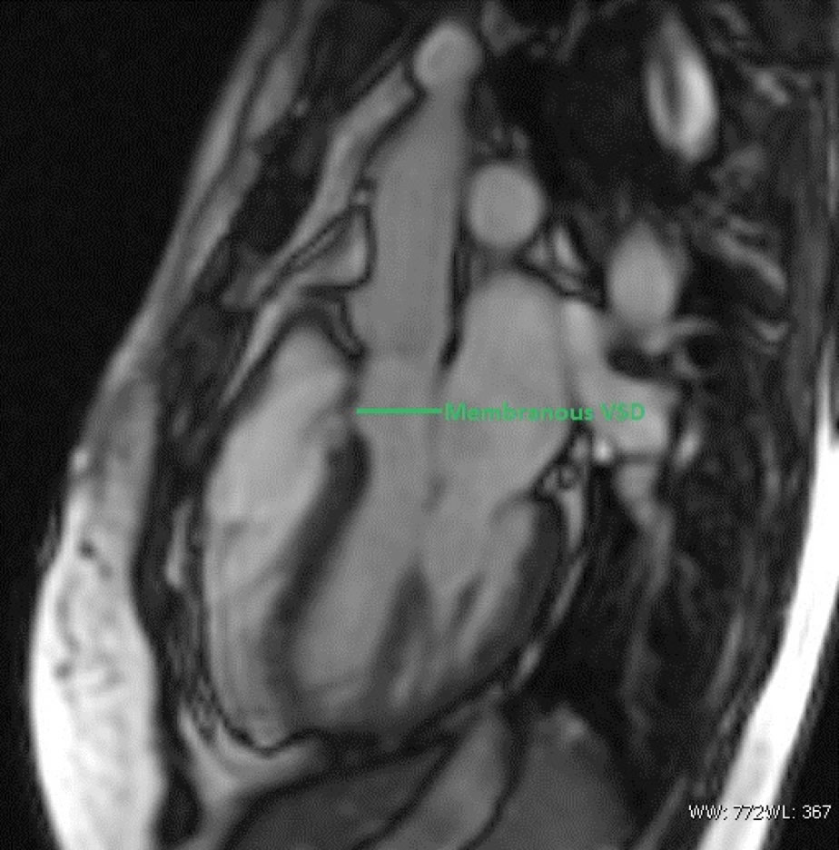
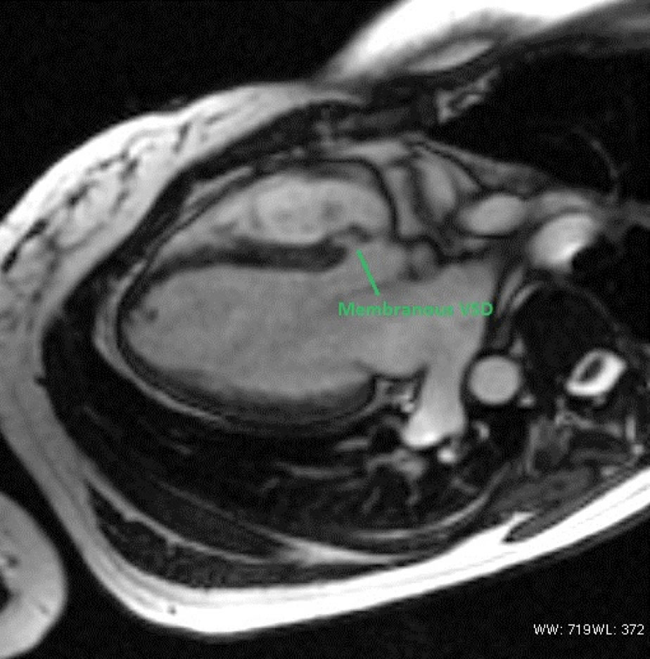
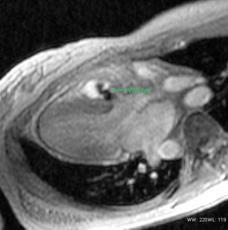
Short and long-axis images are used to visualize VSD and blood shunting.
Short and long-axis images
5. Which imaging findings affect treatment?
The severity of VSD can be quantified in terms of Qp/Qs. Surgery is generally reserved for patients with Qp/Qs > 1.5, unless they are going for cardiac surgery for other reasons.
6. Drawbacks of existing tests:
Echocardiography is most commonly used to assess VSD. It is also used to assess LV size and function. However, it cannot measure the ventricular volumes.

